Case competition 2017のご報告

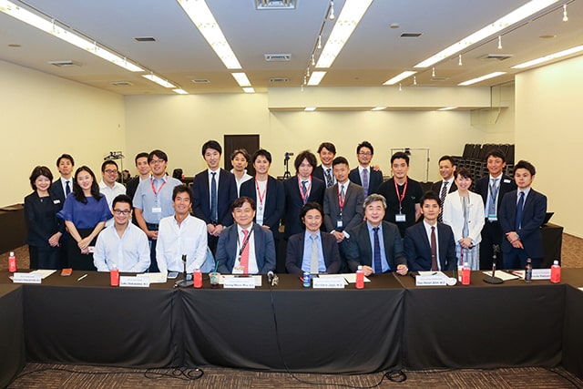
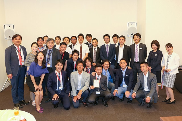
TOPIC2017開催期間に合わせて、第4回SUNRISE Case Competitionを開催致しました。
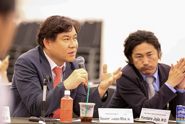
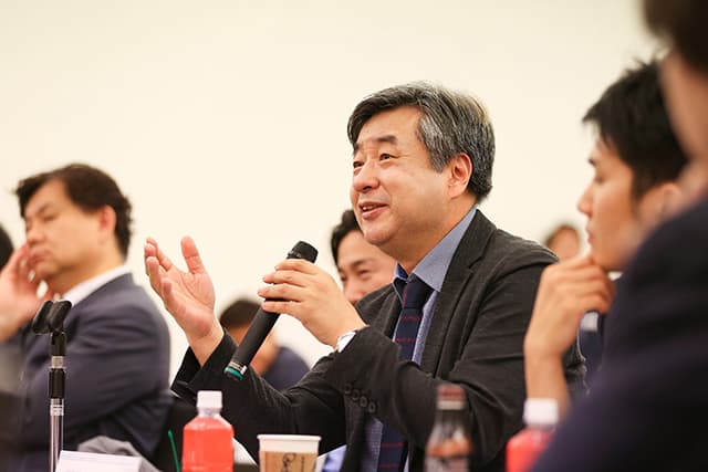
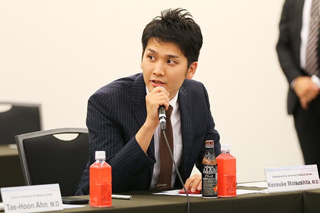
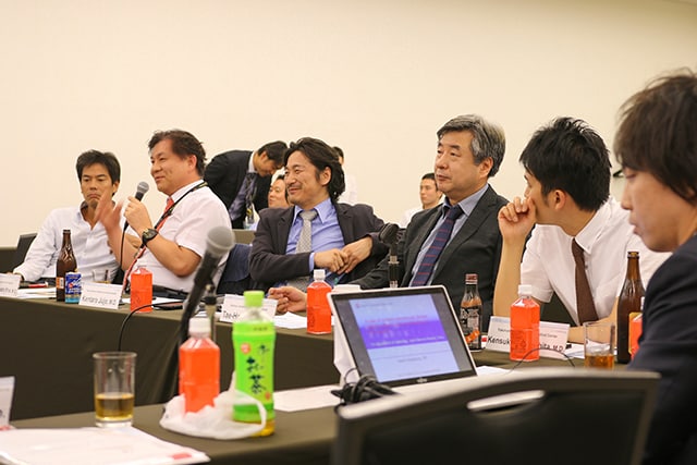
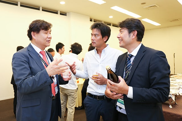
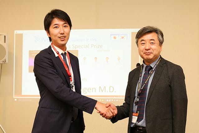
Guest commentatorsとして、例年どおりKoreaからSeung-Woon Rha先生、Tae-Hoon Ahn先生を招聘。さらに前回優勝者の松下絢介先生をjudgeとしてお招きし、TOPIC会場付近の会議室での開催となりました。今年からは冠動脈ありのルールで行われ、2名のrevengerを含む6名のdoctorsにご発表頂きました。年々レベルアップするプレゼンスキルと、coauthorsを巻き込んだ白熱するディスカッションのなか盛会のうちに幕を閉じました。
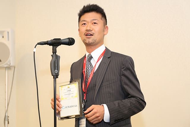
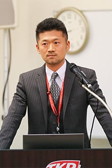
厳正なる採点の結果、『Best Presentation Award』は菱刈(ひしかり)景一先生が受賞されました!この少し強面のヤングライオンには今年のGuro live(10/19-21 Seoul)のinvited facultyとなっていただき、lectureと2nd operatorを務めていただくことになります。
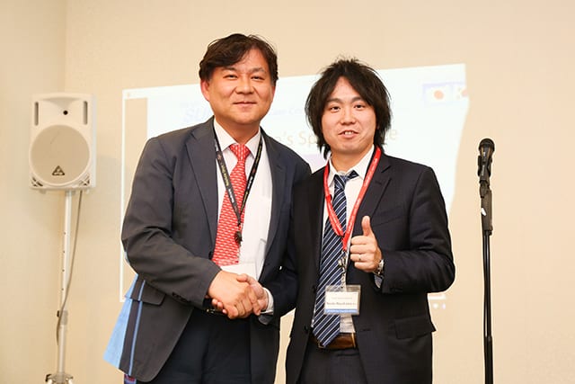
また、1点差で2名のrevengersが特別賞を受賞。早川直樹先生にはGuro liveで、三軒豪仁先生にはEncore liveでfacultyとしてお仕事してきて頂きます。
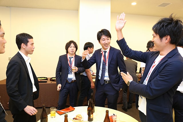
毎回シンデレラボーイが誕生するこの会にまだご参加いただけていない先生は、次回ぜひpresenterもしくはaudienceとしてライブでご覧下さい!
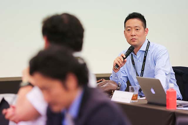
【Presenters】(発表順)
1.松田 裕治 先生 (東京医科歯科大学)
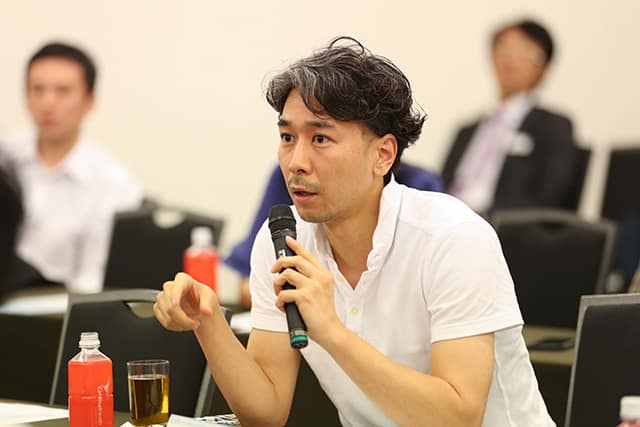
2.三軒 豪仁 先生 (日本医科大学)
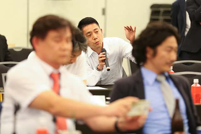
3.中林 圭介 先生 (春日部中央総合病院)
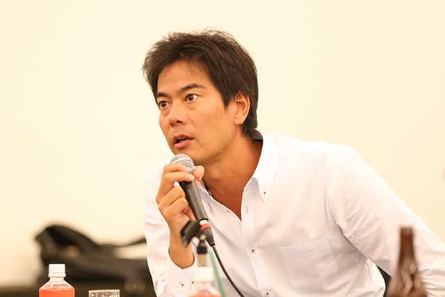
4.山下 雄司 先生 (東京労災病院)
5.菱刈 景一 先生 (横須賀共済病院)
6.早川 直樹 先生 (旭中央病院)
【Best Presentation Award】
菱刈 景一 先生 (横須賀共済病院)
 A Novel Endovascular Strategy for Treating Chronic Total Occlusion of the Aortoiliac Artery Using the ACUNAVTM Ultrasound Catheter
Purpose: Endovascular recanalization of chronic total occlusion (CTO) of aortoiliac artery is technically challenging. A significant proportion of procedural failure has resulted from limited anatomical information with conventional imaging tools. Recently, a monoplane real-time intravascular ultrasound catheter (IVUC, AcuNavTM; Siemens Medical Solutions, Inc., Mountain View, CA) has been developed. We report a patient with iliac CTO who was endovascularly treated with AcuNavTM IVUC.
Case Report: The patient was 63-year-old woman with a total occlusion of left common iliac artery to common femoral artery. During endovascular therapy, duplex ultrasound could not get clear images of the culprit lesion due to severe calcification of the plaque, obesity, and excessive bowel gas. Conversely, AcuNavTM IVUC that was placed in the intra-iliac vein obtained clear images of the CTO lesion. As a result, we successfully passed the wire through long CTO lesion using the all-stage monitoring function of the AcuNavTM catheter.
Conclusions: In our experience, the procedure under the guidance of real-time AcuNavTM intravascular ultrasound catheter is one of the promising options of successful endovascular therapy for CTO lesions in aortoiliac arteries.
【Special Presentation Award1】
早川 直樹 先生 (旭中央病院)
 A case of complex polyvascular disease: efficacy of the combination of EVT and PCI
A 57-year-old male visited to our institution complaining dyspnea on effort. Echocardiography showed low left ventricular ejection fraction (LVEF) as 30%, and coronary angiography did chronic total occlusion (CTO) both in the left anterior descending (LAD) and right coronary artery (RCA). Furthermore, aortography revealed total occlusion from juxta renal aorta to bilateral common femoral artery, known as Leriche syndrome. This patient was at very high risk for receiving coronary artery bypass grafting or aortic bypass surgery. Thus, we decided to perform combination of percutaneous peripheral and coronary intervention (PCI). At first, we deployed five S.M.A.R.T. stents by kissing stenting technique. Final aortography revealed appropriate stent expansions of aortoiliac lesion. Then, we moved to elective PCI to CTO in RCA. Because there were collaterals from each coronary artery and they were jeopardized, we decided to perform PCI via antegrade route, rather retrograde approach with very high risk. Finally, we could pass the guidewire through CTO lesion using IVUS guided parallel wire technique, and three EES were successfully deployed to occluded RCA. We have learned from this case that the combination of PCI and endovascular therapy was one of the feasible options to treat patients with polyvascular disease and reduced LVEF.
【Special Presentation Award1】
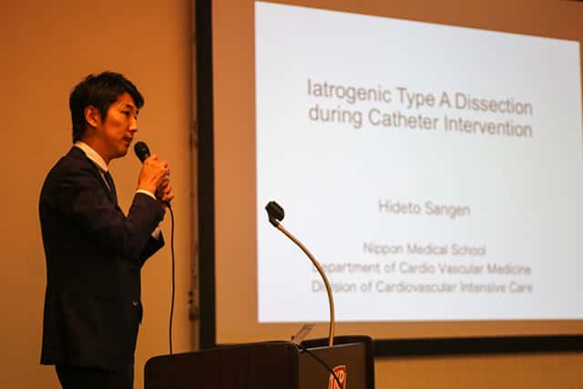
三軒 豪仁 先生 (日本医科大学)
 Iatrogenic Type A Dissection during Catheter Intervention
Background: Iatrogenic type A aortic dissection is one of the severe complications of percutaneous coronary intervention (PCI).
Case: A 43-year-old women was admitted to our hospital due to acute coronary syndrome. Emergent coronary angiogram (CAG) showed total occlusion of posterior descending artery (PDA), subsequently, PCI was performed. When we injected the contrast through guiding cath after a guidewire passed the occluded lesion, the patient cried with pain. CAG revealed ascending aortic dissection occurred. Intravascular ultrasound (IVUS) also showed an intracoronary dissection from ostium to mid portion of right coronary artery (RCA) and the entry point at the proximal RCA. Coronary artery dissection was considered to extend both antegradely and retrogradely. To cover the dissection flap and stop progression of dissection immediately, we attempted IVUS-guided coronary stenting. A Xience Alpine (4.0 x 23 mm) was deployed from ostium to mid portion of RCA to cover the distal end and the entry point of the dissection. All procedures were done without contrast injection. Deployed stent seemed to fully cover the coronary dissection on a final angiogram. Enhanced computed tomography (CT) immediately after the procedure revealed that intramural hematoma had extended to the aortic arch without any contrast enhancement. Finally, coronary CT angiogram on day 17 showed there was no dissection remained in RCA and aortic dissection had been restored completely. The patient discharged on day 20.
Conclusion: IVUS-guided stenting without contrast injection for iatrogenic type A aortic dissection was feasible in this case.