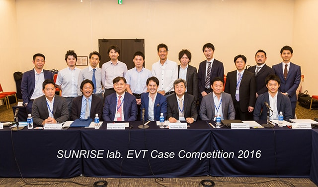
EVT Case Competition 2016のご報告

TOPIC2016開催に合わせて、第3回EVT Case Competitionを開催致しました。
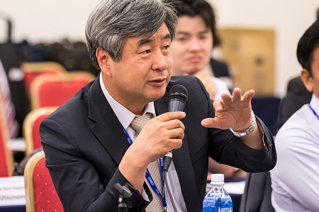
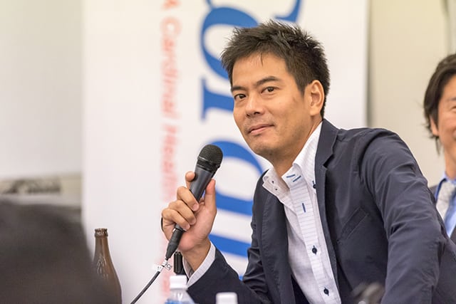
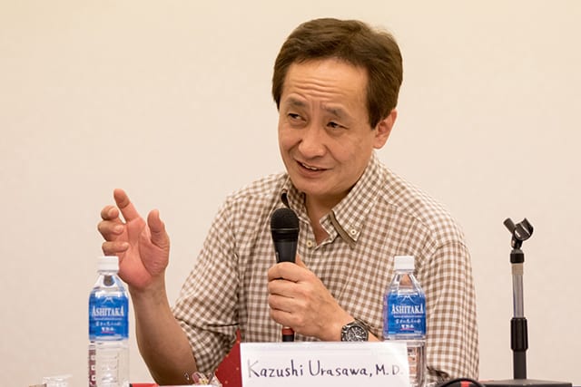
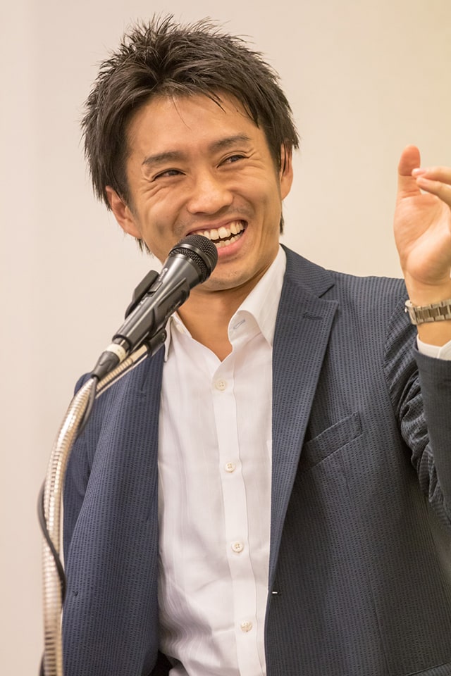
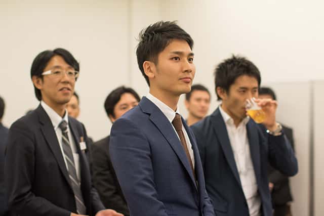
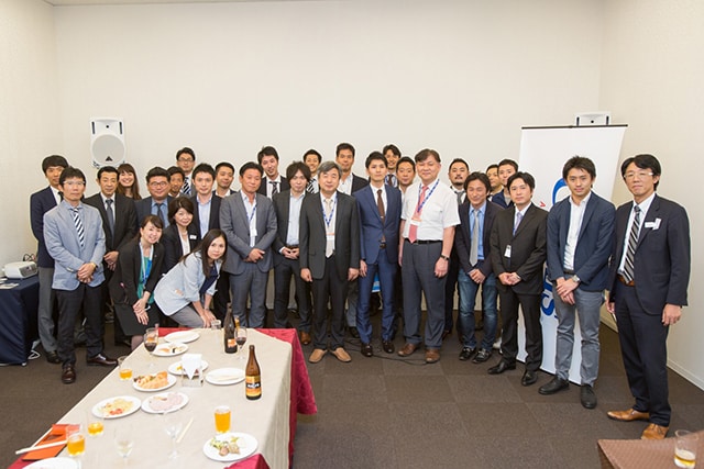
Guest commentatorsとして、例年どおりKoreaからSeung-Woon Rha先生、Tae-Hoon Ahn先生を招聘。また今回は新企画として、前回受賞者の山口徹雄先生と相原英明先生をjudgeとしてお招きし、TOPIC会場付近の会議室での開催となりました。さらにサプライズゲストとして浦澤一史先生に飛び入り参加(!)していただいたこともあり、2名のrevengerを含む7名のdoctorsによるプレゼンテーションは、予定時間を大幅にオーバーして盛会のうちに幕を閉じました。
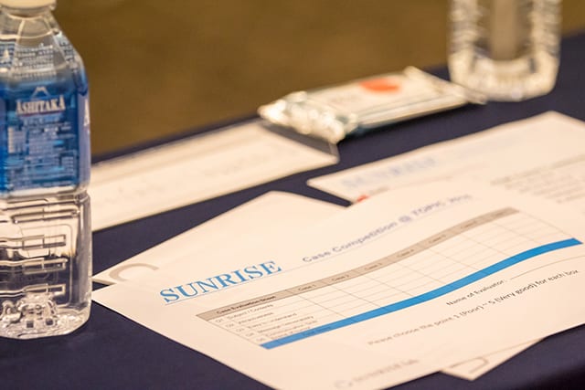
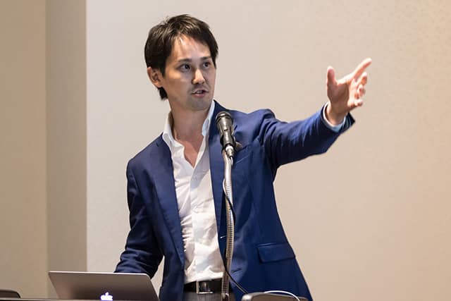
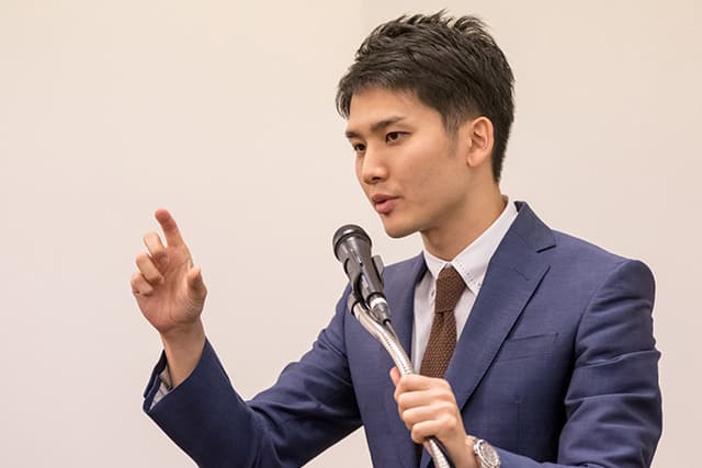
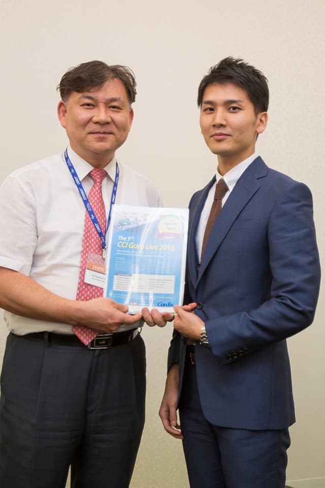
のべ7名のjudgesによる採点の結果、『Best Presentation Award』は松下絢介(けんすけ)先生が受賞されました!この英語が達者なヤングライオンには今年のGuro live(10/27-29 Seoul)のinvited facultyとなっていただき、lectureと2nd operatorを務めていただくことになります。
毎回giant killingが起こるこの会にまだご参加いただけていない先生は、次回ぜひpresenterもしくはaudienceとしてライブでご覧下さい!
【Presenters】(発表順)
1.仲間 達也先生(宮崎市郡医師会病院)
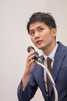
2.松下 絢介 先生(横浜市立大学附属市民総合医療センター)
3.寺村 真範 先生(一宮西病院)
4.椿本 恵則 先生(京都第二赤十字病院)
5.三軒 豪仁 先生(日本医科大学)
6.御手洗 敬信 先生(聖マリアンナ医科大学横浜市西部病院)
7.早川 直樹 先生(旭中央病院)
以下は見事prize getterとなられた松下先生のabstractです。ぜひご覧あれ。
【Best Presentation Award】
A rare cause of claudication
松下 絢介 先生(横浜市立大学附属市民総合医療センター)
 Abstract
A 66-year-old male presented with worsening claudication in the right lower extremity limiting his activities. His right ankle-brachial index (ABI) was 0.50 and left ABI was 1.23. A faint pulse could be felt in his right popliteal artery. Computed tomography (CT) revealed total occlusion at the right popliteal artery and duplex ultrasound showed external compression of the popliteal artery by hypoechoic lesions. Angiography revealed severe stenosis of the popliteal artery and good collateral arteries. Intravascular ultrasound (IVUS) clearly showed iso-hypoechoic lesions between the media and adventitia compressing the true lumen of the popliteal artery and he was diagnosed with cystic adventitial disease (CAD). In order to improve his symptom, low pressure balloon angioplasty was performed. His right ABI improved to 1.08 after the treatment and he was free from symptom at the time he discharge from our hospital. Three years after the treatment, CT revealed a patent right popliteal artery and a residual low density area surrounding the popliteal artery. CAD is a rare cause of claudication which is usually diagnosed by CT, magnetic resonance imaging, and angiography. This case indicated the utility of IVUS to diagnose CAD.